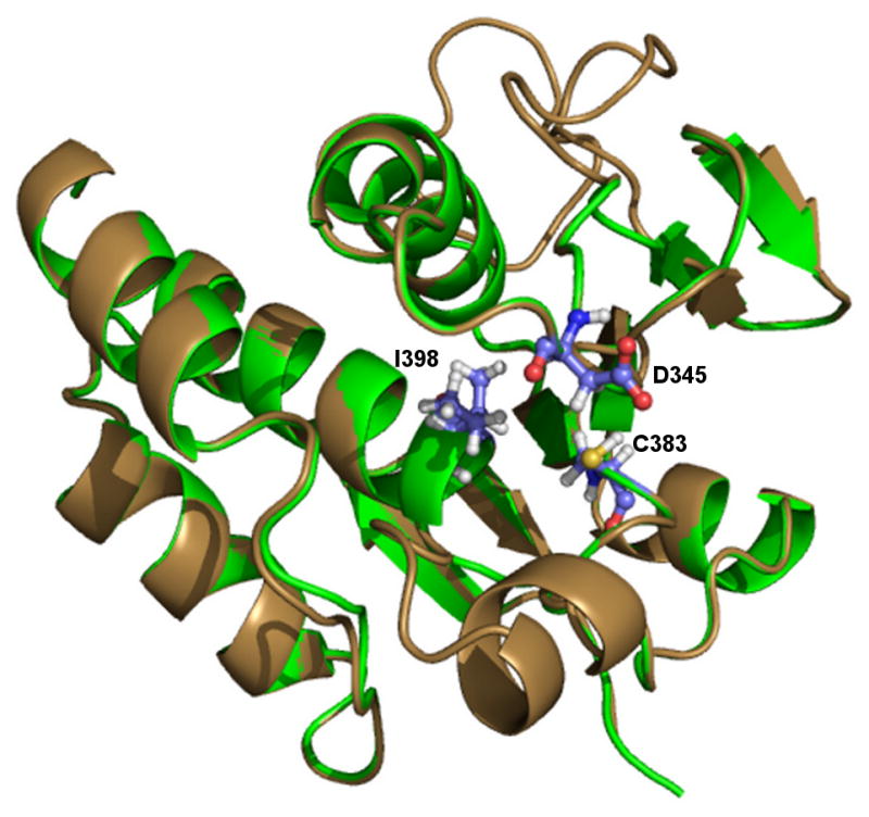
Figure 3.

Homology model of PF3D7_1305500. The structure of PF3D7_1305500 (brown) was developed using the crystal structure of MKP3 as a template (PDB 1MKP). Alignment of the model with MKP3 (green) revealed that the final structure showed the catalytic residues aligned to the proposed positions in the active site. The presence of the signature motif insertion does not affect the shape of the active site and forms an alpha-helix adjacent to the binding pocket without obstructing the site.
