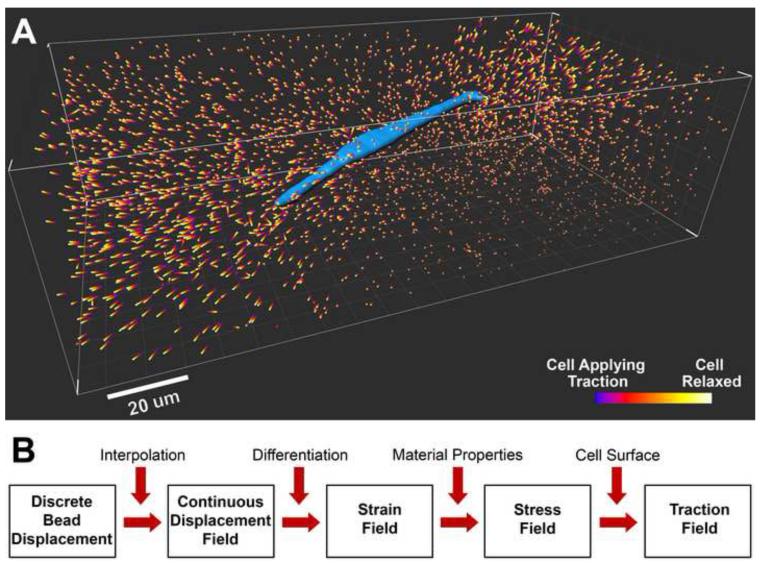
Figure 1. Discrete 3D bead displacements around a cell and forward computation algorithm for mapping cell traction.
A. A breast tumor cell (blue, MDA-MB-231 cell line) is embedded in a type I collagen matrix with concentration of 2 mg/ml. The colored bars are bead displacements caused by the relaxation of the cell after treatment with cytochalasin D. B. Flow chart of a forward computation algorithm to translate bead displacements into a 3D traction field on the cell surface.

