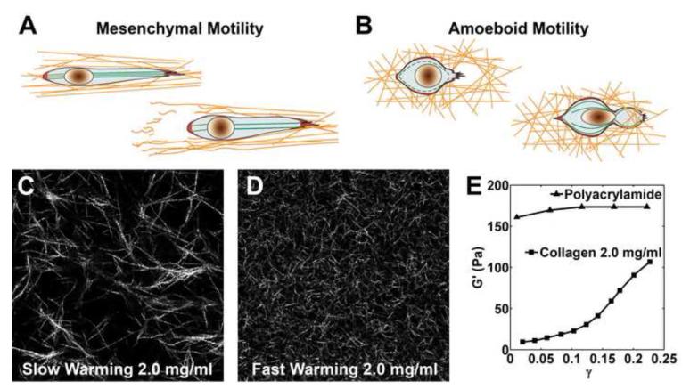
Figure 2. Cellular Motility Modes and Fibrous Nonlinear Collagen Gel.
A. Mesenchymal Motility. Strong traction at the front and back of the cell aligns surrounding fibers as the cell migrates in a protease-dependent manner leaving degraded collagen fibers in its wake. B. Amoeboid Motility. The cell exerts short lived tractions distributed over its surface and squeezes through a pore in a protease-independent manner. (Both A and B are reproduced from Pathak et al.[7] with permission of the Royal Society of Chemistry.) C –D. Confocal reflectance microscopy images of C slow warming and D fast warming type I collagen gel at 2 mg/mL. Image size is 100 um ×100 um. Increasing warming rate during polymerization dramatically decreases pore size and fiber diameter altering the local and bulk mechanical properties of the gel. E. A collagen gel (2 mg/mL) exhibits strain hardening as its shear storage modulus G’ increases with shear strain γ. This is in contrast to the linearity exhibited by a polyacrylamide gel which has constant G’ at varying γ. (The shear storage moduli G’ for both gels was measured at 10 rad/s with a strain-controlled rheometer (RFS-II Rheometrics). The shear storage modulus G’ approaches the shear modulus G at low frequency. Data is reproduced from Storm et al.[73] with permission of Nature Publishing Group.)

