
Figure 1.
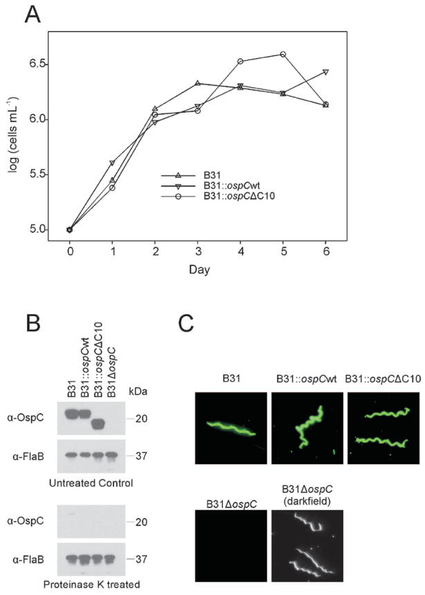
Characterization of wild type and mutant B. burgdorferi strains. In panel A growth rates were determined by daily triplicate cell counts using dark field microscopy of cultures grown at 37°C in BSK-H medium (no antibiotics). In panel B the production and surface presentation of OspC by each strain was assessed by immunoblotting and proteinase K treatment, respectively. Proteinase K treated or untreated cells were fractionated by SDS-PAGE, immunoblotted and screened with polyclonal anti-OspC (phyletic type A) or anti-FlaB antiserum. Surface presentation was further demonstrated through indirect immunofluorescence assays using polyclonal anti-OspC antiserum (Panel D). All methods are detailed in text.