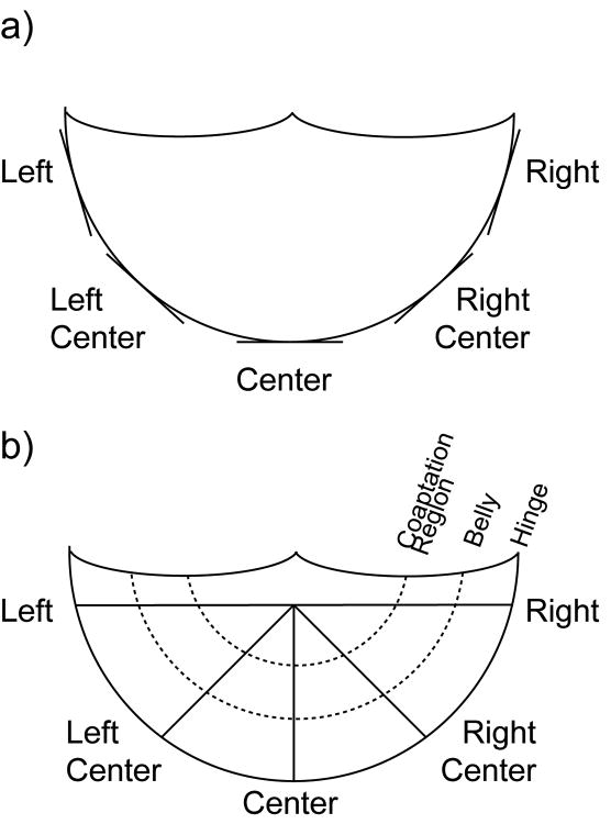
Figure 1.
Schematic of sections cut for IHC and histology. Circumferential sections were cut along the annulus. Radial sections were cut from the center of the free edge to the annulus. Within radial sections, the elastic fiber structure in three regions was analyzed: hinge, belly, and coaptation region.

