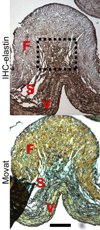
Figure 5.
Elastin IHC (elastin=brown, cell nuclei=purple) and Movat's pentachrome stains (collagen-yellow, PGs/GAGs=blue, elastin=black, cell nuclei=purple) of a collagen fold in the belly, taken from a center section. Whereas the spongiosa was thin through the majority of the belly, underneath collagen folds such as this one, elastin emanated from the ventricularis and branched out to connect the fibrosa. Scale bar=100 μm.

