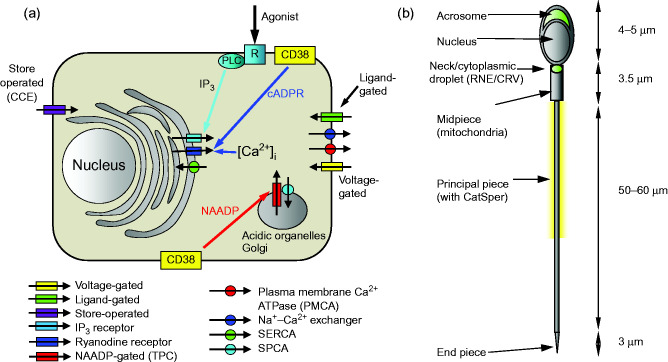
Figure 1.
(a) Simplified diagrammatic summary of [Ca2 +]i signalling toolkit in a somatic cell. Ion channels are shown as rectangles with arrow indicating normal direction of Ca2 + flow (yellow, voltage-gated; green, ligand-gated; purple, store-operated; light blue, IP3 receptor; dark blue, ryanodine receptor; red, NAADP-gated). Pumps are shown as circles with arrows indicating normal direction of Ca2 + movement (red, PMCA'; blue, Na+–Ca2 + exchanger; green, SERCA; blue, SPCA). Activation of IP3 receptors by membrane receptor activation and phospholipase C is shown in light blue. Generation of cADPR and NAADP by CD38 and possibly other enzymes (leading to mobilisation of Ca2 + from intracellular stores) is shown by yellow boxes. (b) Structure of human sperm showing positions of CatSper channels (yellow shading around anterior flagellum) and Ca2 + stores in the acrosome and at the sperm neck (redundant nuclear envelope and calreticulin-containing vesicles) (shown in green).

 This work is licensed under a