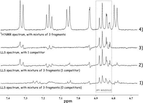
Figure 4.

Identification of a weak binder in a mixture. 1) Weak LLS signals of the spy ligand after sustaining the LLS for Δ=2.5 s in the absence of a competing binder in mixture 1 (spy ligand [II]=500 μm with KD=790 μm protein [Hsp90]=2.5 μm and three nonbinding ligands: 600 μm tyrosine, 600 μm 3,4-difluorobenzylamine, and 600 μm 4-trifluoromethylbenzamidine). 2) Enhanced LLS signals in the presence of a weak binder (mixture 2 contains 600 μm of the weakly binding ligand [V] 3-bromo-5-methylpyridin-2-ylamine (KD=2.2 mm), instead of 600 μm of the nonbinding ligand 3,4-difluorobenzylamine). 3) LLS signals observed in the presence of only the binding fragment (mixture 3 contains 500 μm spy ligand [II], 2.5 μm protein [Hsp90], and 600 μm of the weakly binding ligand [V] 3-bromo-5-methylpyridin-2-ylamine). 4) Conventional 1H NMR spectrum of mixture 2.
