
Figure 7.
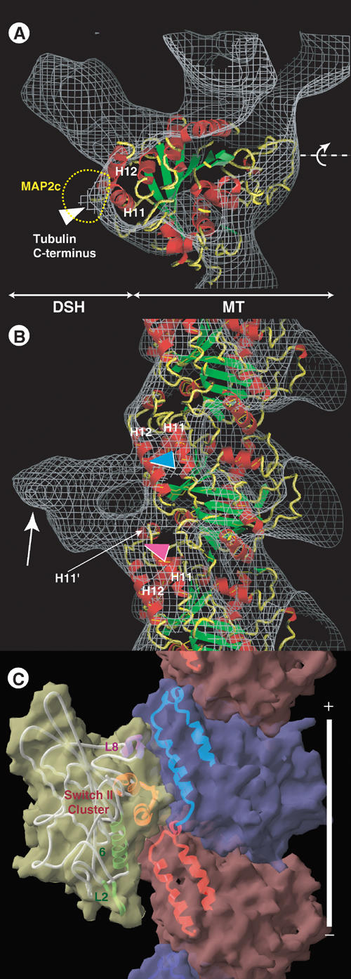
Docking of the tubulin structure onto the EM-derived structure of the DSH–microtubule. (A) Superimposition of the EM-derived map (gray chicken-wire surface) of the DSH–microtubule complex combined with the atomic model of tubulin (ribbon diagram). (B) Side view. Several elements of the motor and tubulin structure are specified to facilitate orientation. In (A), the dashed yellow line indicates the MAP2c/tau outer shape. (C) Side view of the atomic models of the KIF1A–microtubule complex in an ATP-like state.