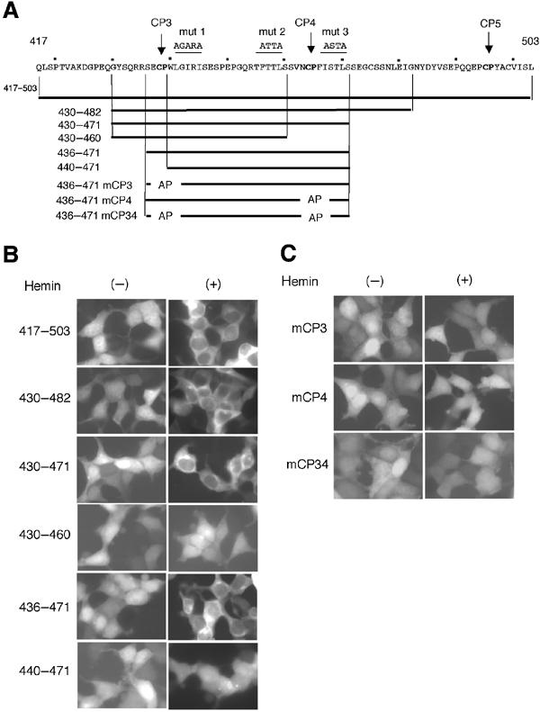
Figure 7.

Demarcation of heme-regulated NES. (A) Amino-acid sequence (residues 417–503) containing CP3–5 motifs (bold) is shown. Mutations in hydrophobic residues are shown above the sequence (mut 1, 2, and 3). Subfragments examined as EGFP fusion proteins are shown below the sequence. (B, C) Indicated EGFP fusions were expressed in 293T cells and their distribution was observed in the absence or presence of hemin.
