Figure 3.

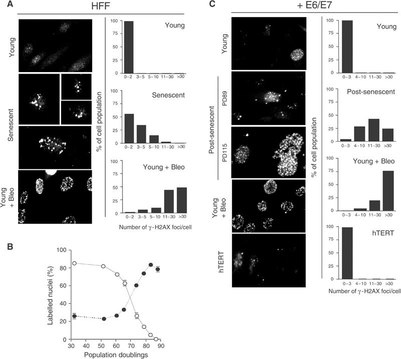
Accumulation of phosphorylated H2AX (γ-H2AX) foci in senescent and E6/E7-expressing fibroblasts grown beyond senescence. (A, C) Immunofluorescence images of fibroblasts (HFF) and fibroblasts expressing E6/E7 (+E6/E7) immunostained with anti-γ-H2AX antibodies. (A) Young at PD39 and senescent at PD82 fibroblasts. (C) Young E6/E7 cells at PD43, post-senescent E6/E7 cells at PD89 and PD115 and immortalised through ectopic expression of hTERT (hTERT). Treatment of young cells with bleomycin is indicated (young+bleo). Right panels show quantification of γ-H2AX foci. The number of γ-H2AX foci per nucleus was counted for each sample and nuclei were categorised as indicated. Note that quantification shown for post-senescent E6/E7 cells refers to cells at PD115. Panels are representative of four independent experiments. (B) Age-dependent increase in the incidence of γ-H2AX foci in HFF cultures. The percentage of cells with γ-H2AX foci (closed symbols) versus the percentage of cells incorporating BrdU (open symbols) is shown at the indicated PD. Bars: standard deviation.
