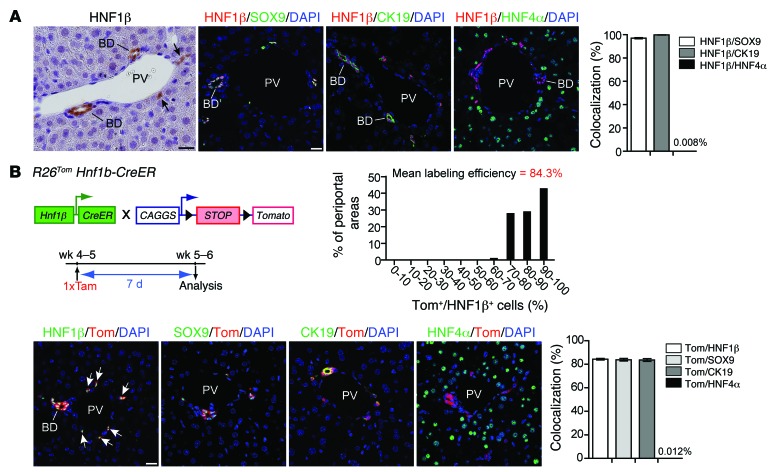
Figure 1. Efficient and specific inducible lineage labeling of the biliary compartment in R26Tom Hnf1b-CreER animals.
(A) In the adult mouse, HNF1β was specifically expressed in bile ducts and biliary ductules/canals of Hering (arrows). HNF1β+ cells were identical with SOX9+ and CK19+ cells, as assessed by co-IF (HNF1β+/SOX9+: 97.04% ± 0.52%, n = 3; HNF1β+/CK19+: 99.89% ± 0.10%, n = 5), while HNF1β expression in HNF4α+ hepatocytes was virtually absent (0.008% ± 0.004%, n = 8). (B) Cre-induced expression of the fluorescent dye tdTom was determined in 5- to 6-week-old R26Tom Hnf1b-CreER reporter mice 7 days after a single tamoxifen injection. tdTom expression was restricted to HNF1β+/SOX9+/CK19+ bile ducts and periportal ductules (n = 5), whereas labeling of HNF4α+ hepatocytes was a rare event (0.012% ± 0.003%, n = 10). All periportal areas revealed a high tdTom labeling efficacy of the HNF1β+ compartment (>30 portal tracts per mouse were quantified at ×600 magnification from 3 discontinuous sections; mean labeling efficiency: 84.3% ± 0.6%; n = 5 animals). BD, bile duct; PV, portal vein; Tam, tamoxifen. Scale bar: 20 μm.

