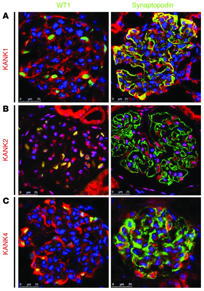
Figure 2. KANK1, KANK2, and KANK4 localize to podocytes in rat glomeruli.

(A) Coimmunofluorescence of KANK1 with podocyte marker proteins in rat glomeruli. KANK1 is highly expressed in podocytes, as identified by the expression of nuclear WT1. KANK1 partially colocalizes with synaptopodin. (B) Coimmunofluorescence of KANK2 with podocyte marker proteins in rat glomeruli. KANK2 is expressed in cytoplasm and nuclei of podocytes. KANK2 does not colocalize with synaptopodin. (C) Coimmunofluorescence of KANK4 with podocyte marker proteins in rat glomeruli. KANK4 localizes to podocyte cell bodies and primary processes but does not colocalize with synaptopodin. Scale bar: 25 μm.
