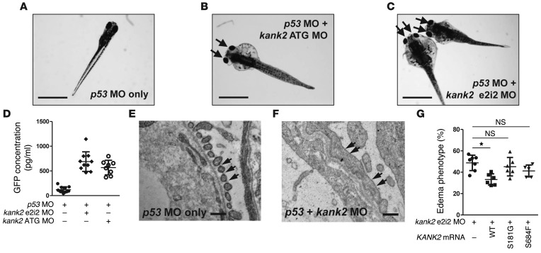
Figure 4. Functional analysis of kank2 knockdown in zebrafish.
(A) Control zebrafish injected with p53 MO. p53 MO did not produce any phenotype until 168 hours after fertilization (n > 100). (B) Zebrafish coinjected with a MO targeting the translation initiation site of zebrafish kank2 (ATG MO) and p53 MO. At 120 hours after fertilization, kank2 morphants display the nephrosis phenotype of periorbital edema (arrows) and body edema in 54.5% of embryos (113 of 207). (C) Zebrafish coinjected with a MO targeting exon 2 splice donor site of kank2 (e2i2 MO) and p53 MO. e2i2 MO causes edematous phenotypes (arrows) in 52.3% of embryos (243 of 464). (D) Proteinuria assay in I-fabp::VDBP-GFP transgenic zebrafish. Note that knockdown of kank2 by either e2i2 or ATG MO causes significant proteinuria compared with that in control fish. (E and F) Electron microscopic structure of glomerular basement membrane and podocyte foot processes in 5-day-old (E) control and (F) kank2 morphant zebrafish. In the control, the foot processes are regularly spanned by slit diaphragms (arrows). In contrast, the foot processes of morphants are effaced and disorganized, with only occasional intercellular junctions (arrows in F). The glomerular basement membrane is disorganized. (G) Functional analysis of KANK2 mutations in zebrafish. Coinjection of kank2 e2i2 MO with a human wild-type KANK2 mRNA (156 of 491) partially rescued edematous phenotypes of kank2 morphants, whereas injection of KANK2 mRNA bearing the p.S181G (277 of 623) or p.S684F (187 of 447) mutation failed to rescue the phenotype. Error bars indicate the SD of more than 3 independent experiments. *P < 0.05, 2-tailed Student’s t test. Scale bar: 1 mm (A–C); 1 μm (E and F).

