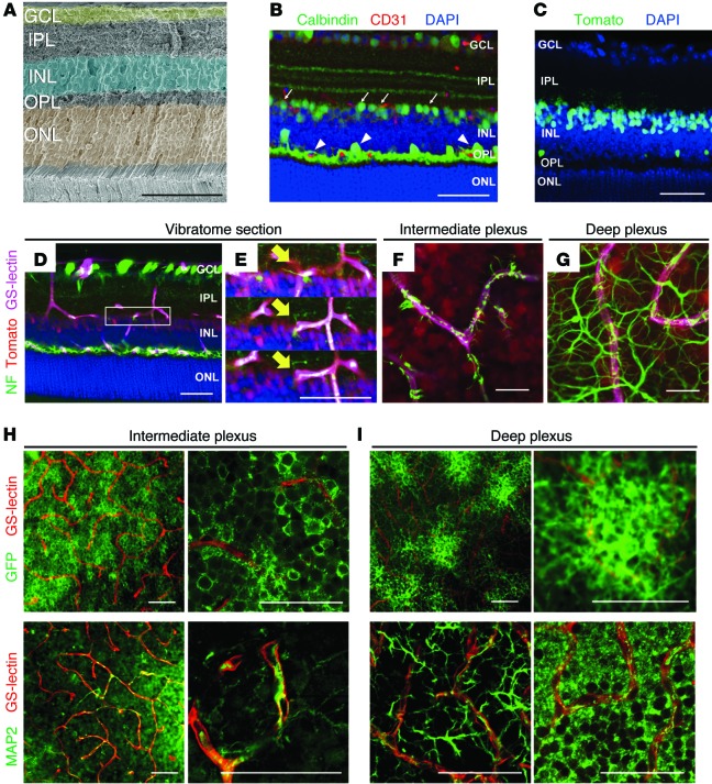
Figure 1. Amacrine and horizontal cells form neurovascular units with the intraretinal capillaries.
(A) Pseudocolored cross section of an adult murine retina. (B) Immunohistochemistry was used to identify putative neurovascular units between amacrine cells (arrows) or horizontal cells (arrowheads) with the vasculature using anti-calbindin (green), anti-CD31 (red), and DAPI (blue) in WT retinal cryosections at P23. (C) Cre recombination reporters label amacrine and horizontal cell nuclei in P23 Ptf1a-Cre R26tdTomato/+ mice (tomato signal was pseudocolored green). (D–I) Amacrine (D–F) and horizontal cell (D and G) neurites (NF-M labeled, green) associate with the intraretinal vasculature (GS-lectin, blue) as seen in thick cut (100 μm) sections (amacrine/horizontal nuclei, red). (E) Adjacent optical slices from the region of interest boxed in (D); arrows mark colocalization. (F and G) Flat-mounted P23 Ptf1a-Cre R26tdTomato/+ retinas colabeled with anti-neurofilament and GS-lectin (endothelial cell marker). (H and I) Amacrine cell neurites are decorated with GFP in Ptf1a-Cre R26GFP mice and can be observed in close proximity to GS-lectin–positive endothelial cells. Immunofluorescence for MAP2 in whole-mount retinas at P23 also reveals colocalization of amacrine and horizontal cell neurites with the intraretinal vasculature. Scale bars: 50 μm (A–E, H, and I); 20 μm (F and G).

