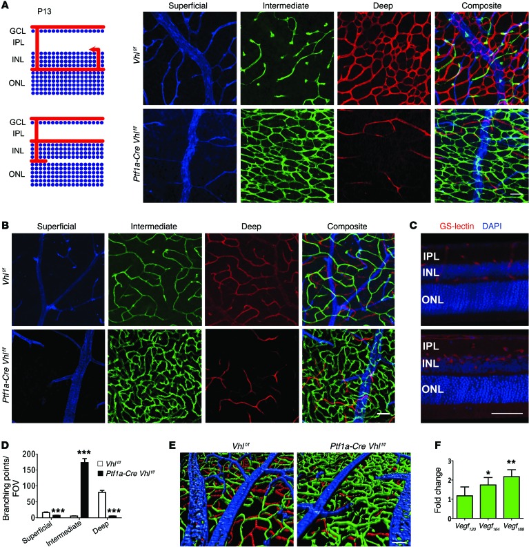
Figure 3. Vhl deletion in amacrine and horizontal cells induces formation of a dense and convoluted intermediate plexus at the expense of the deep plexus.
(A and B) Schematic of angiogenesis in Vhlf/f (control) or Ptf1a-Cre Vhlf/f retinas at P13. Note dramatic alterations in the intermediate plexus (green) and deep plexus (red) at P13 (A) and P23 (B) in flat-mounted retinas. (C) 100 μm sections from P23 Ptf1a-Cre Vhlf/f mice were stained with GS-lectin to highlight the extent of the neovascularization in the VHL mutants. Counterstained with DAPI. (D) The number of branching events in P13 Vhlf/f or Ptf1a-Cre Vhlf/f retinas was plotted (n = 4). (E) Three-dimensional reconstruction of 3 retinal plexuses in P23 Ptf1a-Cre Vhlf/f retina (superficial, blue; intermediate, green; deep plexus, red) highlighted the abnormally dense intermediate plexus. (F) qPCR analyses revealed that nondiffusible Vegf188 was the most abundant isoform expressed in Ptf1a-Cre Vhlf/f mice at P15 (n = 4). *P < 0.05, **P < 0.01, ***P < 0.001; 2-tailed Student’s t tests. Error bars indicate mean ± SD. Scale bars: 50 μm (A–C and E).

