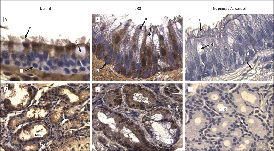
Figure 4.
Serum amyloid A2 immunoreactivity in the sinus mucosa followed by hematoxylin counterstaining. A and D, Control tissues. B and E, Chronic rhinosinusitis (CRS) tissues. C and F, Negative controls. A-C, Epithelia. D-F, Glands. Original magnification ×400 for all panels. Ab indicates antibody; b, basal cell; c, ciliated cell; g, goblet cell; and smg, submucosal gland.

