
Figure 3.
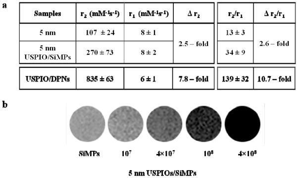
Relaxometric characterization of the magnetic nanoconstructs. a, The transversal (r2) and longitudinal (r1) relaxivities, and the r2/r1 ratio are listed for the free USPIOs and the corresponding USPIO-loaded nanoconstructs, as derived from a bench-top relaxometric analysis. The change in transversal relaxivity Δr2 and Δr2/r1 ratio is also provided, showing a significant enhancement in MRI performance for all configurations. Note that for the DPNs, the enhancement is calculated with respect to the hydrophilic 5 nm USPIOs. b, Phantom images for different concentrations of the 5 nm USPIO-loaded SiMPs (magnetic nanoconstructs) generated using a 3T Philips MRI clinical scanner. Note that 107 SiMPs are equivalent to ~ 0.2 μg of Fe.