Fig. 3.
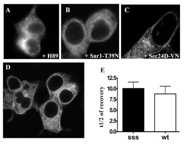
Accumulation of GAT1-SSS in punctate structures requires the COPII-dependent ER-export machinery. (A) HEK293 cells were transfected with a plasmid encoding YFP-tagged GAT1-SSS. Cells were treated overnight with H89 (100 μM). Images were acquired 24 hours after transfection. (B,C) HEK293 cells were co-transfected with plasmids coding for CFP-tagged GAT1-SSS (1 μg) and Sar1-T39N (2 μg, B) or YFP-tagged Sec24D-VN (2 μg, C). Images were acquired 24 hours after transfection. (D) HEK293 cells were transfected with a plasmid coding for the double mutant YFP-tagged GAT1-RL/AS-SSS and images were captured 24 hours after transfection. (E) HEK293 cells were co-transfected with plasmids encoding SAR1A-T39N (2 μg) and either YFP-tagged wild-type GAT1 (wt; 1 μg) or YFP-tagged GAT1-SSS (sss; 1 μg). FRAP was recorded as outlined in the Materials and Methods. Data represent mean half-lives in seconds of fluorescence recovery from five independent experiments; error bars indicate s.e.m.

