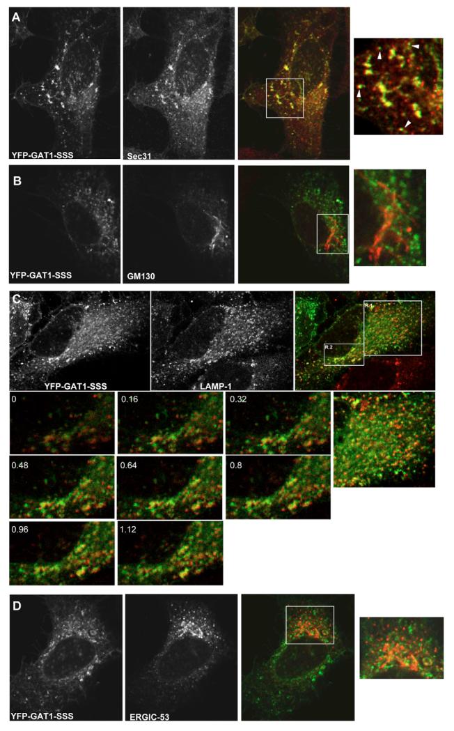
Fig. 4.
Colocalization of GAT1-SSS with subcellular markers. HeLa cells were transfected with plasmids encoding YFP-tagged GAT1-SSS. After 24 hours, cells were fixed and immunostained against the ERES marker Sec31 (A), the cis-Golgi marker GM130 (B), the late endosome/lysosome marker LAMP1 (C) and a marker for the intermediate compartment, ERGIC53 (D). (A,B,D) Boxed areas are shown as close-ups on the right. (A) Arrowheads highlight GAT1-SSS punctae that are closely associated with ERES. (C) The numbered images represent magnifications of region 2 (R.2), which were acquired in multiple optical slices. The numbers indicate how many μm the optical slices are from 0. To the right of these is a close-up of region 1 (R.1). Images were acquired using a confocal microscope (Leica SPE).

