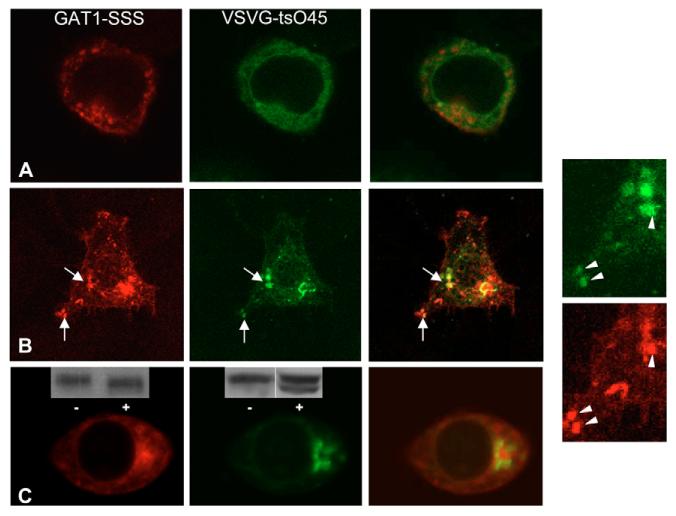
Fig. 6.
Transient colocalization of YFP-tagged VSVG-ts045 with CFP-tagged GAT1-SSS during its trafficking through the anterograde secretory pathway. HEK293 cells were co-transfected with plasmids encoding CFP-tagged GAT1-SSS and YFP-tagged VSVG-ts045. Cells were incubated for 20 hours at 40°C. Images were acquired using a confocal microscope before (A) and at 10 (B) and 30 (C) minutes after shifting to the permissive temperature. The two images on the right show close-ups of those in B. (C) Insets show immunoblots of CFP-tagged GAT1-SSS (left-hand panel) and YFP-tagged VSVG-ts045 (middle panel): cell extracts were prepared at 30 minutes after shifting to the permissive temperature and incubated overnight in the absence (−) or presence (+) of endoglycosidase H. Immunoreactive material was visualized by an antiserum directed against GFP.

