
Figure 6.
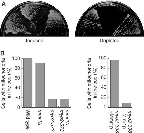
Effect of Mmr1p loss on myo2 mutants. (A) pGAL1-MMR1:mmr1 (left sector), myo-338 pGAL1-MMR1:mmr1 (middle sector), and myo2-573 pGAL1-MMR1:mmr1 (right sector) cells were streaked on SCRGD (left) and SC (right) plates, and incubated at 25°C for 3 days. (B) Left: cells with the indicated genotype (siblings from the cross between mmr1Δ and myo2-573) were grown to mid-log phase at 25°C in SC. Right: myo2-338 pGAL1-MMR1:mmr1 cells were grown to mid-log phase in SCRGD (myo2-338+Mmr1p), or shifted to SC and incubated for 3 h (myo2-338−Mmr1p) at 25°C. Cells were stained with DASPMI, and budded cells were counted (n>200).