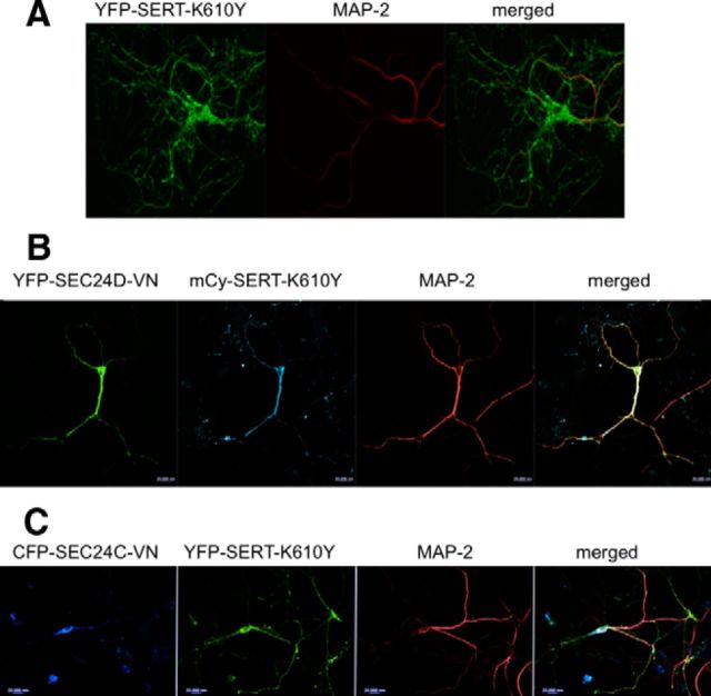
Figure 7.
Axonal targeting of YFP-tagged SERT–K610Y, an SEC24D client. Rat dorsal raphe neurons were transfected with plasmids encoding YFP–SERT–K610Y (A), YFP–SEC24D–VN and mCherry–SERT–K610Y (mCy-SERT-K610Y; plasmid ratio of 5:1; B), or CFP–SEC24C–VN and YFP–SERT–K610Y (plasmid ratio of 5:1; C) using Lipofectamine2000. After 48 h, the neurons were fixed and stained for MAP-2. MAP-2 staining was detected using Alexa Fluor 568- or Alexa Fluor 633-conjugated secondary antibodies. Images were captured by confocal microscopy. YFP-tagged SERT–K610Y reached the MAP-2-negative compartment in 14 of 14 examined and in 15 of 15 examined neurons in the absence and presence of CFP–SEC24C–VN. During coexpression of dominant-negative SEC24D, mCherry-tagged SERT–K610Y was confined to the MAP-2-positive compartment in 16 of 20 neurons. Data are from four independent experiments (i.e., 4 individual preparations of rat dorsal raphe neurons that were independently transfected in parallel with a plasmid coding for a tagged version of SERT–K610Y and plasmids encoding CFP–SEC24C–VN or YFP–SEC24D–VN). Scale bars, 20 μm.

