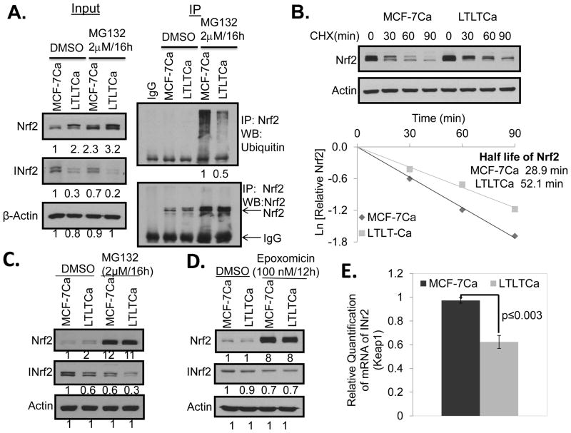
Figure 3. LTLTCa cells showed decreased ubiquitination and degradation of Nrf2 compared with MCF-7Ca cells.
(A) 1 mg of total cells lysate from MG-132 treated cells was immunoprecipitated with 1 μg of rabbit IgG or Nrf2 antibody. The immunoprecipitated Nrf2 was immunoblotted for ubiquitin and Nrf2. (B) Cells were treated with 25 μg/ml cycloheximide (CHX) for the indicated time points and 30 μg of total cells lysate was immunoblotted with Nrf2 and β-actin antibodies. The graphs represent the natural logarithm of the relative levels of the Nrf2 protein versus the CHX chase time and the half-life of Nrf2 was determined using the linear part of the degradation curve. LTLTCa cells showed no difference in rate of INrf2 protein degradation but demonstrated lower levels of INrf2 transcripts, as compared with MCF-7Ca cells. (C) Cells treated with MG-132, and (D) with epoxomicin were lysed and immunoblotted. (E) Total RNA was isolated from the cells and cDNA was synthesized from 1 μg of total RNA and the cDNA was used to quantify the INrf2 gene transcripts at basal level.

