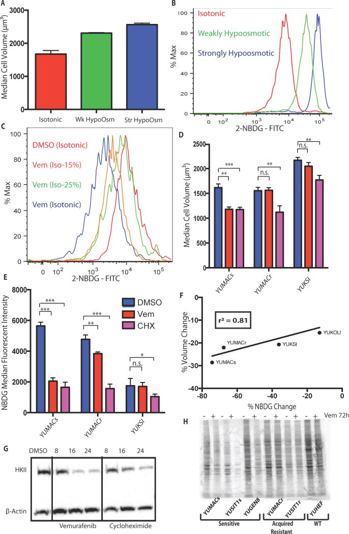
Figure 6.
Glucose uptake is greatly affected by changes cell volume. A, Decreasing solution osmolarity while incubating with 2-NBDG causes both an increase in cell volume and B, 2-NBDG uptake that is osmolarity-dependent. The isotonic condition has a total concentration of 289 mOsm/L, while the weakly and strongly hypoosmotic conditions have concentrations of 144.5 mOsm/L and 72.25 mOsm/L respectively. C, Incubation of 3μM vemurafenib-treated cells in hypoosmotic 2-NBDG solution restored glucose uptake to the same level as isotonic 2-NBDG control at 217 mOsm/L. D. Inhibition of protein translation with 50 μg/mL cycloheximide induces a decrease in both cell volume and E, glucose uptake at 24 hours with F, a relationship similar to 3μM vemurafenib (r2 = 0.8153, p = 0.097). G, Inhibition of translation with cycloheximide produces comparable loss of HKII to Vemurafenib by 24h. H, 35S methionine labeling shows that incubation of vemurafenib sensitive but not resistant lines for 72h with 3μM reduced total new protein synthesis, *, p < 0.05; **, p < 0.01; ***, p < 0.001.

