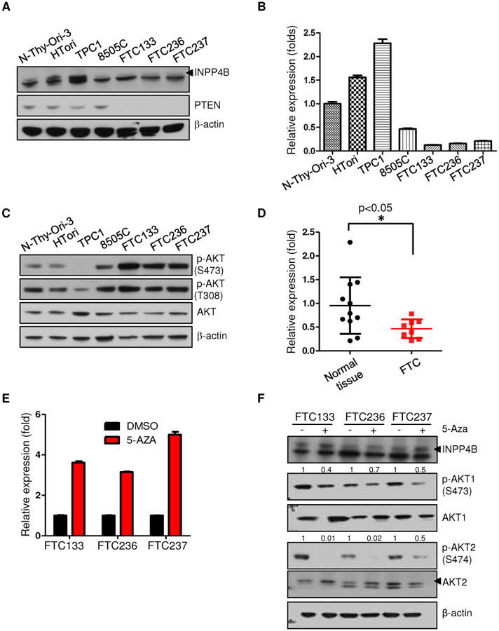
Figure 3. INPP4B expression is reduced in human follicular thyroid cell lines and human surgical specimens.

A. Western blot analysis of thyroid cancer cell lines for INPP4B and PTEN expression. Arrow indicates specific band. The arrowhead indicates the specific band of INPP4B protein. B. RT-qPCR analysis of thyroid cancer cell lines for INPP4B transcript levels. C. Western blot analysis of thyroid cancer lines for AKT activation on both Serine 473 and Threonine 308 residues. D. RT-qPCR analysis of INPP4B in unmatched normal versus FTC patient tumor samples. E-F. Thyroid cancer cell lines were treated with 3uM 5-Aza-2′-deoxycytidine for 5 days. Panel shows transcript (E) and protein analysis of INPP4B expression and AKT activation levels in DMSO versus 5-Aza treated FTC cells. (F). Arrows indicate specific band (see also Methods). AKT2 antibody used is (5239).
