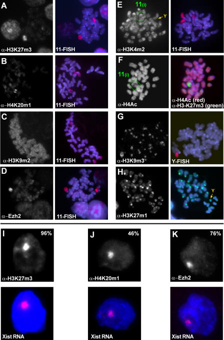
Figure 1. Epigenetic Imprints at the Initiation of X Inactivation.
(A–H) Indirect immunofluorescence and subsequent DNA FISH analysis on mitotic chromosomes prepared from undifferentiated clone 36 ES cells after 3 d of Xist induction. H3K27m3 (A), H4K20m1 (B), and Ezh2 (D) are enriched on the arms of Chromosome 11 upon ectopic Xist expression. H3K9m2 (C) is not enhanced upon Xist expression. H3K4m2 (E) is reduced on Chromosome 11 upon Xist induction (green box) and absent from pericentric heterochromatin and the Y chromosome (orange arrow). (F) Histone H4 multiple-lysine acetylation is partially reduced (green box, left panel). Hypoacetylation (red) is restricted to chromosomal regions which show high levels of H3-K27 trimethylation (green, right panel). H3K9m3 (G) and H3K27m1 (H) are enriched at constitutive heterochromatin of pericentric regions and the Y (orange arrows).
(I–K) Indirect immunofluorescence (upper panels) and subsequent Xist RNA FISH (red, Xist RNA; blue, DAPI) analysis of H3K27m3 (I), H4K20m1 (J), and Ezh2 (K) in interphase nuclei of undifferentiated clone 36 ES cells expressing Xist for 3 d.

