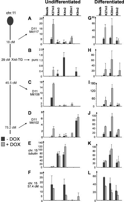
Figure 2. ChIP Mapping of H3K27m3, H4K20m1, H3K9m2, H3K4m3, and H3K4m2 on the Xist-Expressing Chromosome 11 during Differentiation of Clone 36 ES Cells.
A genetic map of Chromosome 11 indicating the loci analysed is given on the left (Xist-TG, approximate integration site of Xist transgene; puro, PGKpuromycin marker).
(A to F) Chromatin was prepared from undifferentiated clone 36 ES cells grown for 3 d in the presence (light bars) or absence (dark bars) of doxycycline. H3K27m3 and H4K20m1 were enriched at three intergenic microsatellite sequences at 18.0 (A), 45.5 (C), and 75.2 (D) cM. (B) H3K27m3 was established over the coding sequence of PGKpuromycin in doxycycline-induced cells, which was accompanied by a loss of H3K4m2 and H3K4m3. (E) Tubulin control. (F) Control microsatellite located on Chromosome 15.
(G–L) Analysis of H3K27m3, H4K20m1, and H3K9m2 in clone 36 ES cells differentiated for 9 d with (light bars) or without (dark bars) doxycycline. Histone methylation marks were monitored. Experiments were performed in duplicate, and the standard error is indicated in the graphs.

