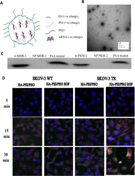
Figure 1.

Characteristics of HA derivative/siRNA particles. (A) Schematic representation of HA-PEI/PEG/EGF nano-assemblies with negatively charged siRNA. (B) The self-assembling nanoparticles showed a spherical morphology in TEM. (C) Electrophoretic retardation analysis of siRNA binding by HA-PEI with the release of intact siRNA by poly(acrylic acid). (D) Cell uptake studies in SKOV-3 WT and TR model. HA-rhodamine-123 and scramble siRNA labeled with fluorescein were incubated with the cells for 5, 15 and 30 min. The cells showed uptake within 15 min of incubation with the dual targeted system showing higher fluorescence signal compared to the CD44 targeted system. The cell nuclei were stained with DAPI (blue).
