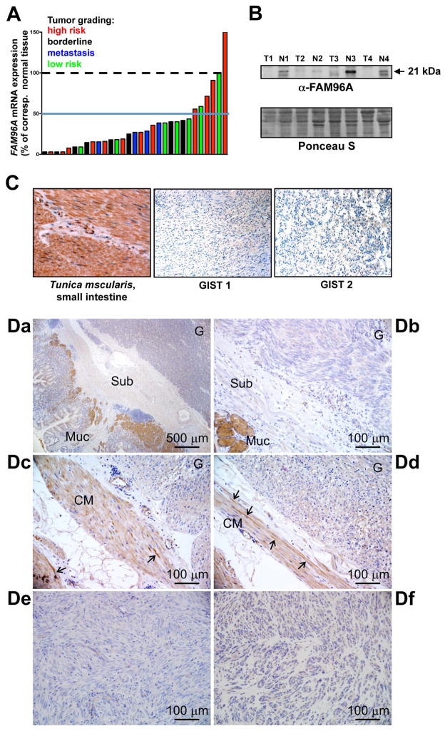
Figure 3.
FAM96A expression is decreased in GIST. (A) FAM96A mRNA in 31 gastric GISTs (21 frozen and 10 paraffin-embedded samples) was compared to 14 normal stomach mucosal samples obtained from the same patients by real-time RT-PCR. Expression data were normalized to the average expression value of the normal tissues. 29 of the 31 tumors showed reduced FAM96A mRNA irrespective of tumor grading (green bars: low risk, blue: borderline, red: high risk, black: metastasis). (B) Western blot analysis of FAM96A protein expression in four pairs of GISTs (T) and corresponding normal samples (N). The tumor samples showed reduced FAM96A expression. (C) FAM96A immunohistochemistry performed in a tissue microarray containing 53 individual GIST samples using a self-raised antibody. Brown color indicates FAM96A immunoreactivity in the tunica muscularis of the small intestine. The complete loss of FAM96A immunoreactivity in a representative spindle-cell (GIST 1) and an epithelioid GIST (GIST 2) is shown at high magnification (200x). (D) FAM96A immunohistochemistry performed using the commercial antibody HPA040459 (see Supplementary Table 2 for patient and sample information). Da, GIST-2; Db, GIST-16; Dc-d, GIST-6; De, GIST-3; Df, GIST-18. Note no or very low FAM96A immunoreactivity in the tumors (labeled G in Da-d); strong staining in mucosal glands (Muc in Da-b), weaker staining in circular smooth muscle cells (CM in Dc-d) and strong staining in interstitial cells (arrows in Dc-d). Sub, submucosa.

