Figure 6.
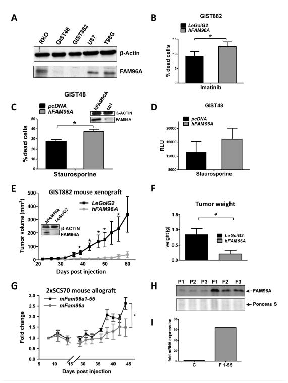
Re-expression of FAM96A in GIST and related cell lines increases apoptosis sensitivity and inhibits tumorigenicity. (A) FAM96A protein expression in GIST and other cancer cell lines detected by Western blotting. GIST48, imatinib-resistant GIST cells; GIST882, imatinib-sensitive GIST cells; U87 and T98G, glioblastoma cells; β-actin, loading control. Both GIST lines displayed severely reduced levels of endogenous FAM96A expression. (B) GIST882 cells lentivirally transduced either with hFAM96A or the empty vector, LeGoiG2, were incubated with 10μM imatinib mesylate for 24 hours. Re-expression of FAM96A was confirmed by immunoblotting (see Figure 6E, inset); cell death was assessed by FCM using 7-AAD. *, P≤0.05; n=4. (C) GIST48 cells were transiently transfected with either hFAM96A or the empty vector, pcDNA3.1. FAM96A re-expression was confirmed by immunoblotting (see Fig. 6D, inset). Apoptosis was induced by incubating the cells with 1μM staurosporine for 16 hours. The proportion of dead cells was determined by FCM using PI exclusion. *, P≤0.05; n=3. (D) Caspase-9 activity was measured in hFAM96A- and empty vector-transfected GIST48 cells 16 hours after treatment with 1μM staurosporine using the Promega Caspase-Glo® 9 Assay. n=3. (E) 7.7×10⁶ GIST882 cells lentivirally transduced either with hFAM96A or the LeGoiG2 empty vector were subcutaneously xenografted into NOD/SCID mice. Overexpression of hFAM96A reduced the frequency of successful engraftment from 8/9 to 5/9 and significantly reduced tumor volumes at 39, 43, 47, 50 and 56 days post-injection (P<0.05). Western blot analysis confirmed stable FAM96A protein overexpression in the FAM96A-transduced GIST882 cells before injection into the mice (inset). (F) The weights of hFAM96A-expressing tumors were significantly reduced upon excision at 60 days post-injection (P<0.05). (G) Tumorigenic 2xSCS70 murine ICC-SC cells were transduced with full-length mFAM96A or the deletion mutant mFAM96A aa1-55, which does not bind APAF, and allografted subcutaneously into NCr-nu/nu mice (5×10⁶ cells/mouse, n=6 mice/group). Overexpression of full-length mFAM96A significantly reduced tumor growth (P<0.05). (H) Verification of stable FAM96A protein overexpression by Western blotting in three tumors each derived from either non-transduced parental (P1–P3) or FAM96A-transduced (F1–F3) 2xSCS70 cells. Ponceau S staining was used as loading control. (I) Because FAM96A aa1-55 is not detected by the self-raised anti-FAM96A antiserum used in this study, we demonstrated overexpression of FAM96A aa1-55 mRNA using quantitative real time RT-PCR. C represents a control pool of RNA isolated from 5 non-transduced parental tumors; F1-55 is a pool of RNA isolated from 5 FAM96A aa1-55-transduced tumors.

