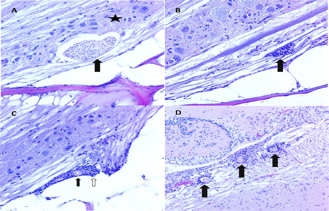
Figure 2. General features of infection.
A. Parasite clusters (PCs) were most commonly observed in the ventral white matter of the spinal cord. In this image, a PC in white matter (arrow) impinges of the gray matter (star). Photomicrograph. H&E. 400× magnification. B. A common feature of infection was myelitis, or inflammation of the spinal cord neuropil. Myelitis in these fish was generally multifocal and composed of inflammatory cells that are most likely a combination of granulocytes and microglial cells (arrow). Photomicrograph. H&E. 400× magnification. C. Meninxitis is inflammation of the perineural membranes of the teleost central nervous system, so called because they do not have a true set of meninges as in mammals. Here, granulocytes (white arrow) surround a ruptured PC (black arrow) at the base of a nerve root. D. Encephalitis, inflammation of the brain, was associated with PCs (black arrows) in the medial longitudinal fasciculus, a descending white matter tract in the rhombencephalon. Photomicrograph. H&E. 200× magnification.

