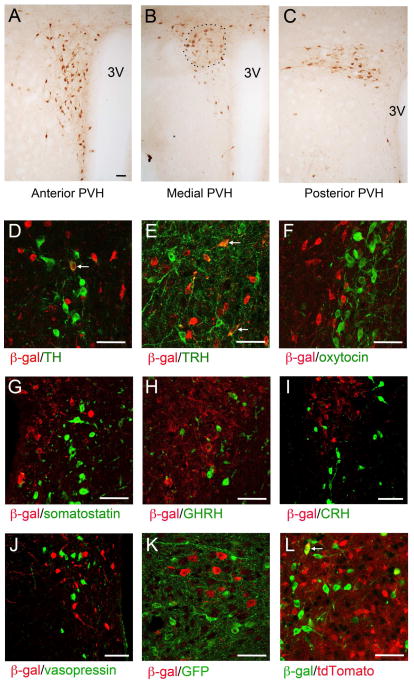
Figure 1. BDNF expression in the PVH as revealed by anti-β-galactosidase immunohistochemistry in BdnfLacZ/+ mice.
(A–C) Immunohistochemistry images showing the distribution of BDNF expression in the PVH. The approximate location of the PVHmpd is outlined. 3V, the 3rd ventricle.
(D–J) Coexpression of BDNF with tyrosine hydroxylase (TH), thyrotropin-releasing hormone (TRH), oxytocin, somatostatin, growth hormone releasing hormone (GHRH), corticotropin-releasing hormone (CRH), and vasopressin in the PVH. BDNF-expressing neurons were marked by β-galactosidase (β-gal) in BdnfLacZ/+ mice. Arrows denote representative BDNF neurons that also express TH or TRH.
(K) Coexpression of BDNF with MC4R in the PVH. BDNF- and MC4R-expressing neurons were marked with β-galactosidase and GFP, respectively, in BdnfLacZ/+;Mc4r-tau-GFP mice.
(L) Coexpression of BDNF with TrkB in the PVH. BDNF- and TrkB-expressing neurons were marked with β-galactosidase and tdTomato, respectively, in BdnfLacZ/+;TrkBCreERT2/+;Ai9/+ mice. The arrow denotes a representative BDNF neuron that also expresses TrkB.
The scale bar represents 50 μm. See also Figure S1.

