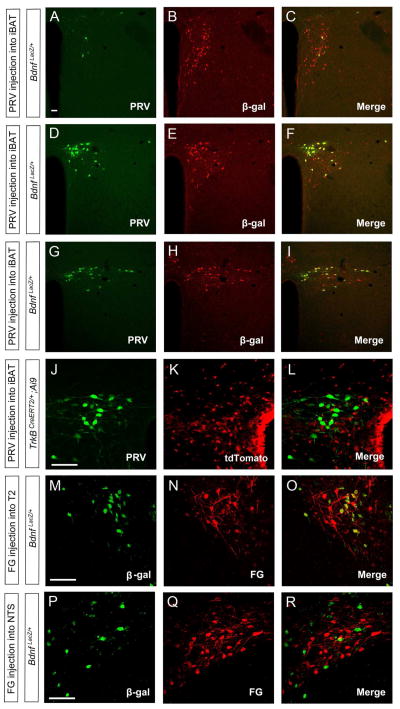
Figure 6. Direct projection of BDNF neurons in the PVH to the spinal cord.
(A–I) Confocal images revealing that some BDNF neurons marked by β-galactosidase (β-gal) were labeled by PRV injected into iBAT in the anterior PVH (A–C), medial PVH (D–F), and posterior PVH (G–I).
(J–L) Representative confocal images showing that PVH neurons labeled by PRV injected into iBAT are distinct from TrkB neurons marked by tdTomato.
(M–O) Confocal images showing that fluorogold (FG) injected into T2 segment of the spinal cord labels many BDNF neurons in the PVH.
(P–R) Confocal images showing that fluorogold injected into the NTS labels only a small number of BDNF neurons in the PVH.
The scale bars represent 50 μm. See also Figures S5 and S6.

