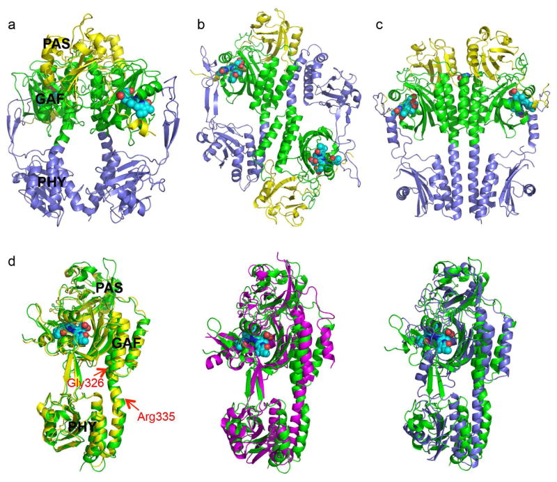
Fig. 1. Crystal structures of RpBphP2 and RpBphP3.
a) Ribbon diagram of the parallel dimer of RpBphP2-Ctag in the Pr state. The PAS (yellow), GAF (green) and PHY (blue) domains are juxtaposed along the dimer interface, where a large void is found. The BV chromophores are shown in cyan spheres. b) Ribbon diagram of the anti-parallel dimer of RpBphP3 in the Pr state. c) Parallel dimer structure of PaBphP in the Pfr state exhibits a tighter dimer interface (PDB ID: 3NHQ). d) Pairwise comparison of the tertiary structures of BphP monomers: RpBphP3 (green; as reference); RpBphP2 (yellow), Cph1 (magenta) and PaBphP (blue). The red arrows mark the locations of kink at the GAF-PHY linker helix (RpBphP3 numbering). Related to Figure S1 and Figure S2a.

