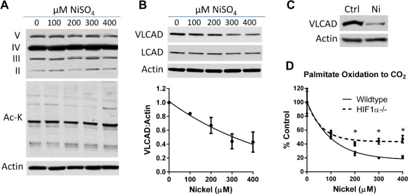
Figure 3. Nickel reduces expression of VLCAD and the effect of nickel on FAO is partially abrogated in HIF1α knockout cells.

(A) The mechanism of nickel sulfate (NiSO4) on FAO does not involve changes to the abundance of the respiratory chain (top panel) or changes in mitochondrial protein lysine acetylation (Ac-K; middle panel). (B) Nickel dose-dependently reduces VLCAD, but not LCAD, protein levels in MEFs. Densitometry from repeated VLCAD blots was used to generate the line graph. (C) Nickel also reduces VLCAD protein abundance in human lung fibroblasts. (D) Wildtype and HIF1α knockout MEFs were treated with the indicated concentrations of nickel for 24 hrs and palmitate oxidation measured. The effect of nickel is significantly blunted at concentrations of nickel >200μM. *P <0.05, HIF1α−/− versus wildtype cells. Assays were performed in quadruplicate.
