. 2015 Jun 29;30(4):434–442. doi: 10.3904/kjim.2015.30.4.434

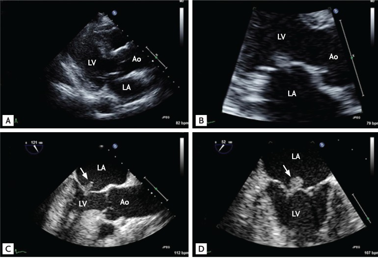
Figure 4. Representative echocardiographic images of a patient with community-acquired infective endocarditis involving an apparently normal-looking valve. The patient, a 66-year-old female with poorly controlled diabetes mellitus, visited the emergency department due to fever and back pain. Blood culture showed Enterococcus faecalis bacteremia. Transthoracic echocardiographic images showed a normal-appearing mitral valve with a suspicious mass attached to the left atrial side of the mitral leaflet (A, B) and transesophageal echocardiography clearly showed a textured surface typical of vegetation (C, D; arrows). Ao, aorta; LA, left atrium; LV, left ventricle.

Figure 4