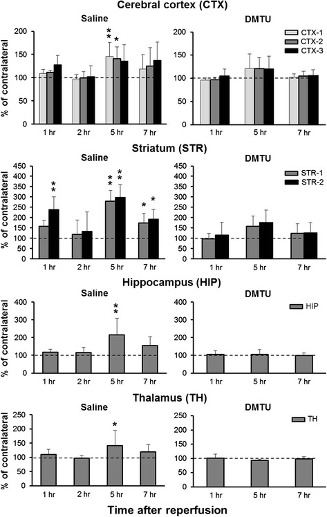
Fig. 3.

Time changes in brain ROS generation detected by [3H]hydromethidine in tMCAO mouse treated with saline and DMTU. [3H]Hydromethidine was intravenously injected into mice treated with saline or DMTU (200 mg/kg, i.p.) 30 min before tMCAO (90 min). Coronal sections at the level of the striatum and hippocampus were prepared at 1, 2, 5, and 7 h after ischemia/reperfusion of MCA in mice. Radioactivity concentrations in the brain including the cortex, striatum, hippocampus, and thalamus were calculated from the autoradiograms based on the ROIs drawn on MRI images (Fig. 2) and are presented as the percentage of the contralateral side. Data are expressed as mean ± SD (n = 5–8 for each time point). *P < 0.05, **P < 0.01, as compared with control mice
