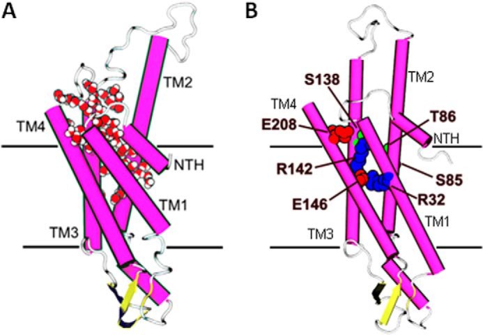
FIGURE 5.

Several sites sensitive to tryptophan substitution face a putative water pocket identified during MD simulations of a Cx26 hemichannel (14). A, one monomer of the Cx26 MD-simulated model showing location of a water pocket accessible from the intracellular face of the membrane. Protein has been colored by secondary structure (magenta, α-helix; yellow, β-sheet; white, loop). Water molecules filling the IC pocket are shown in van der Waals representation colored by element (red, oxygen; white, hydrogen) (Ref. 14 reprinted with permission from Biophys. J.). B, Cx32 model showing only the nonfunctional related residues with hydrophilic properties, red representing negatively charged side chains, blue representing positively charged side chains, and green representing polar side chains.
