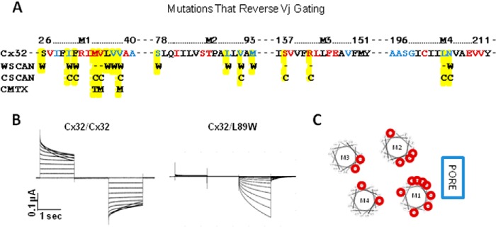
FIGURE 6.

Sites where mutations “reverse” voltage gating in Cx32, possibly through a mechanism that involves disruption of TM domain interactions. A, amino acid sequence of TM domains highlighting tryptophan mutants with abolished (red) or reduced (blue) function. Letters below individual amino acids indicate the substitutions that reverse voltage gating (W, tryptophan mutant; C, cysteine mutant; other, CMTX mutant). Data from Skerrett et al. (33), Toloue et al. (38), and Oh et al. (36). B, junctional currents recorded from oocytes expressing Cx32/Cx32 (left) and Cx32/L89W (right). When recording intercellular currents, both oocytes were clamped at −20 mV, and currents were recorded from a continuously clamped oocyte although its partner was pulsed to +80 and −120 mV in 10-mV steps. C, helical wheel plots arranged to represent the relative positions of TM domains with respect to the channel pore. Sites where point mutations are reverse voltage gating are circled in red.
