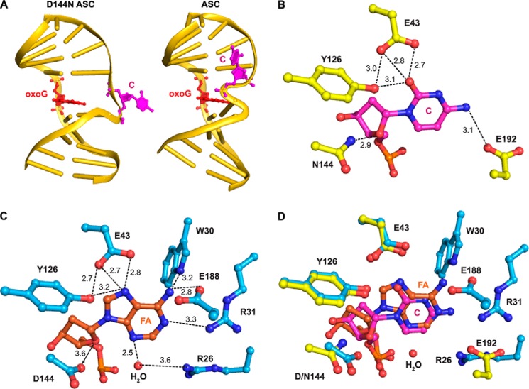
FIGURE 6.
Structure of the D144N ASC. A, global structure of D144N ASC and ASC. The protein portion is not displayed, but DNA that is bound to it is. DNA in both structures is shown in gold, oxoG is shown in red, and C is shown in magenta. The two structures are viewed from the same orientation. B, close-up view of the active site of the D144N ASC. C, close-up view of the active site of the FLRC. D, an overlay of B and C. Protein in FLRC is shown in cyan, 2′-β-fluoroadenosine (FA) is shown in orange, and ordered water is shown as red spheres. Protein in D144N ASC is shown in yellow, and cytosine is shown in magenta. Hydrogen-bonding interactions are denoted in dotted lines, with the associated numbers, showing distances in Å.

