FIGURE 7.
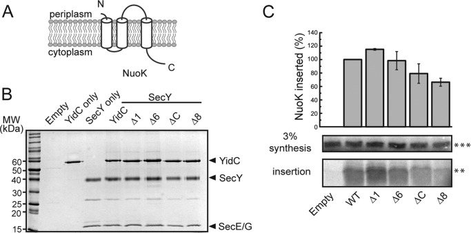
Double deletions interfere with YidC functioning in the membrane insertion of NuoK. A, a topology diagram of E. coli endogenous substrates NuoK. B, protein profiles of co-reconstituted YidC and SecYEG into E. coli liposomes. YidC and SecYEG were co-reconstituted at the molar ratio of 1:1. C, NuoK insertion was analyzed via the proteinase K protection assay. After in vitro synthesis and proteinase K digestion, the membrane-inserted amount of the proteinase K-resistant bands corresponding to the C-terminal deleted version (**) stimulated by both YidC and SecYEG were probed on 16% Tricine gel. 3% of the synthesis reaction was loaded as a control. Protease-protected NuoK and derived fragments were quantified. The amount of inserted NuoK in the presence of wild-type YidC was set to 100. All the data points shown are obtained from the average of three independent experiments, and calculated standard deviation intervals are shown.

