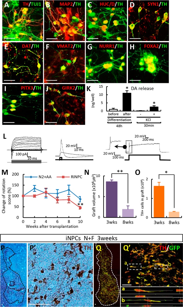
FIGURE 7.
Functional midbrain-like DA neurons are induced from iNPCs in vitro and can be transplanted into a PD rat model. A–D, N+F-transduced iNPCs differentiated over a 10–15-day period. TH+ cells co-expressed neuron-specific markers (TUJ1, MAP2, HuC/D, and synapsin I (SYN1)). E and F, DA homeostasis markers DAT and VMAT2. G and H, exogenously expressed NURR1 and FOXA2. I, TH+ cells expressed proteins specific for mDA neurons (PITX3). J, A9 type midbrain marker GIRK2 co-localized with TH+ cells. K and L, presynaptic DA neuronal functions as seen on electrophysiological analysis (L) and DA release assay (K). DA release was evoked by high potassium-induced depolarization stimulus in differentiated cells. M–Q, in vivo behavior (M–O) and histological analyses (P and Q) after transplantation. Behavioral recovery was assessed by amphetamine-induced rotation in sham-operated animals (N2 + ascorbic acid (AA), n = 6) and NF-induced DA cells (RiNPC, n = 8) (M). Graphs depict the graft volume (N) and total number of TH+ cells in grafts between 3 and 8 weeks post-transplantation (O). At 3 weeks post-transplantation, the presence of GFP-labeled donor iNPCs co-expressed TH+ cells (P, DAB; Q, immunofluorescence image). P′, higher magnification views of P; Q′, region adjacent to Q; a and b, higher magnification views of Q′). Error bars, S.E. *, p < 0.05, compared with the control (K). *, p < 0.05; **, p < 0.001 compared with week 0 (M) and 3 weeks after transplantation (N and O). Scale bar, 50 μm.

