
FIGURE 2.
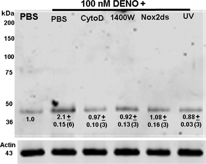
Western blot showing biotinylated proteins. Lysates of neutrophils exposed to PBS + 5.5 mm glucose or to PBS/glucose + 100 nm DENO for 2 min were prepared according to the biotin switch assay. The entire gel is shown. Where indicated cells were concurrently incubation with DENO and 5 μm cytochalasin D, 1 mm 1400W, or 10 μm Nox-2ds. UV indicates cells exposed to UV light for 5 min after DENO incubation. Although not shown, cytochalasin D, 1400W, Nox-2ds, and UV had no effect on biotin switch results in cells incubated with PBS rather than DENO. The numbers beneath the band at ∼43 kDa reflect means ± S.E. (n = replicate experiments) for the fold difference in 43-kDa band density/actin band density normalized to the ratio of PBS-only exposed control cells for each experiment. *, p < 0.05 versus control.