
FIGURE 5.
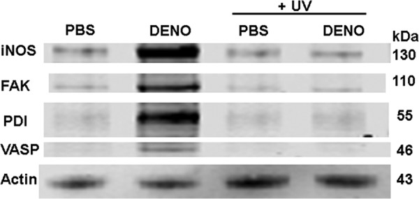
Protein associations in the Triton-soluble F-actin fraction. Murine neutrophils were exposed to PBS/glucose ± 100 nm DENO for 2 min. Where indicated, samples were then exposed to UV light for 5 min prior to addition of DTSP to cross-link proteins, then fractioned based on Triton solubility (see “Experimental Procedures”), and subjected to Western blotting. This is a representative blot among four replicate experiments. Data based on the blots are shown in Table 4.