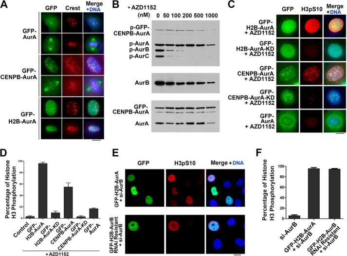
FIGURE 1.
Chromatin-localized Aurora A phosphorylates the Aurora B substrate histone H3 in vivo. A, immunofluorescence labeling of HeLa cells transfected with GFP-Aurora A (AurA), GFP-CENPB-Aurora A, or GFP-H2B-Aurora A with a CREST antibody. The DNA was stained by DAPI. Note that the wild-type Aurora A was localized to the centrosome in interphase, whereas GFP-CENPB-Aurora A was situated to the centromere and GFP-H2B-Aurora A to the chromatin/chromosome both in interphase and mitosis. Scale bar, 10 μm. B, HeLa cells transfected with GFP-CENPB-Aurora A were synchronized to prometaphase and treated with different concentrations of Aurora B inhibitor AZD1152 for 1 h. Cell extracts were subjected to Western blot analysis with antibodies to phospho-Aurora A/-B/-C, Aurora B (AurB) and Aurora A. Note that AZD1152 at 200 nm and above totally inhibited the activation of Aurora B and Aurora C (AurC) indicated by autophosphorylation, whereas 200 nm AZD1152 did not inhibit the activation of GFP-CENPB-Aurora A. C, histone H3 Ser(P)-10 immunofluorescence labeling of HeLa cells transfected with GFP-H2B-Aurora A, GFP-H2B-Aurora A-KD, GFP-CENPB-Aurora A, GFP-CENPB-Aurora A-KD, or GFP-Aurora A followed by treatment with 200 nm AZD1152 for 1 h. The DNA was stained with DAPI. Note that GFP-H2B-Aurora A and GFP-CENPB-Aurora A expression resulted in phosphorylation of, and colocalized with histone H3 Ser-10 (H3pS10), whereas GFP-H2B-Aurora A-KD, GFP-CENPB-Aurora A-KD, and GFP-Aurora A did not induce histone H3 Ser-10 phosphorylation. Scale bar, 10 μm. D, quantitative characterization of HeLa cells treated as in C. Positive staining cells are defined as strongly staining with clear edge. Cells with no stain or weak vague staining are not counted as positive staining cells. Bars represent the percentage of the cells with histone H3 Ser-10 phosphorylation. Each data point represents 3 independent experiments with each measuring 100 cells, and error bars indicate S.D. E, histone H3 Ser(P)-10 immunofluorescence labeling of HeLa cells co-transfected with Aurora B siRNA to knock down endogenous Aurora B and GFP-H2B-Aurora A or RNAi resistant GFP-H2B-Aurora B. Note that both GFP-H2B-Aurora B and GFP-H2B-Aurora A phosphorylated histone H3 Ser-10 (H3pS10). F, quantitative characterization of HeLa cells treated as in E. Bars represented the percentage of the cells with histone H3 Ser-10 phosphorylation. Each data point represents three independent experiments with each measuring 100 cells, and error bars indicate S.D. Scale bar, 10 μm.

