FIGURE 5.
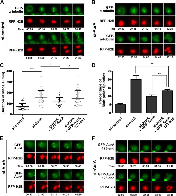
N-terminal deletion of Aurora A results in prolonged mitosis and multipolar spindle assembly. A and B, HeLa cells stably expressing GFP-α-tubulin were transfected with irrelevant (A) or Aurora A (AurA) siRNA (B) and subjected to time-lapse microscopy. RFP-H2B was transiently expressed as a chromatin/chromosome indicator. Note that Aurora A depletion resulted in multipolar cell division. Scale bars, 10 μm. C and D, quantitative characterizations of the duration of mitosis with live cells (C) and percentage of the cells with abnormal spindle assembly with fixed cells (D) shown in (A, B, E, and F) (see below). *, p < 0.05; **, p < 0.01; ***, p < 0.0001. The duration of mitosis is determined from the nuclear envelope breakdown to two daughter cells formation. Each data point represents 3 independent experiments with each measuring 50 cells, and error bars indicate S.D. E and F, HeLa cells co-transfected with Aurora A siRNA-, RFP-H2B-, and RNAi-resistant GFP-Aurora A (E) or GFP-Aurora A aa123-end (F) were viewed by time-lapse microscopy. Note that Aurora A aa 123-end could not rescue Aurora A depletion. Scale bars, 10 μm.

