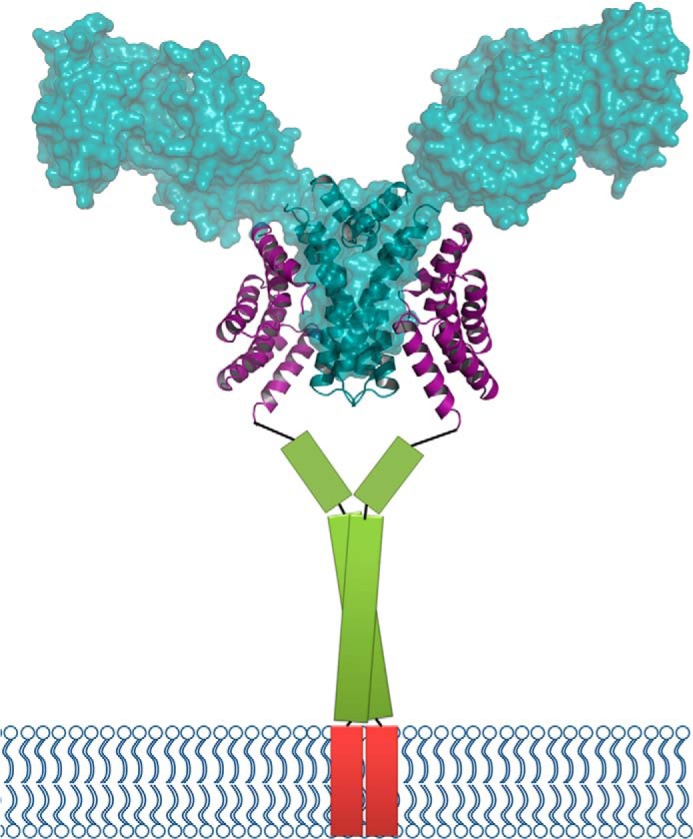
FIGURE 8.

Model of the FLU-GluTR complex. The FLUTPR-GluTRDD complex in ribbon representation is overlaid with the transparent surface of GluTR (PDB code 4N7R, chains A and B). The predicted coiled-coil domain of FLU is shown as a green column, and the transmembrane domain is shown as a red column.
