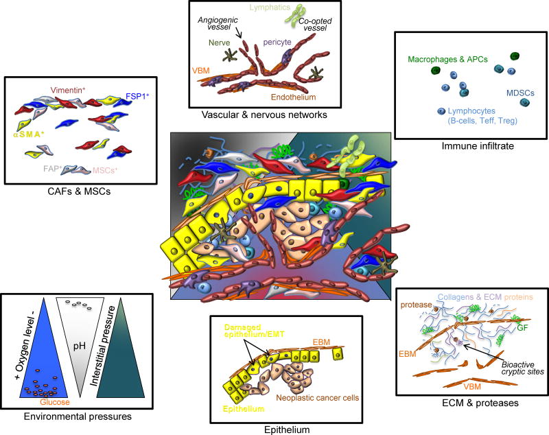
Figure 1. Heterogeneity of the TME.
At the center, an overlay of defined TME components depicts the complexity and heterogeneity of the TME. Cancer associated fibroblasts (CAFs) and mesenchymal stem cells (MSCs) express various proteins often times used for classification; some of which are alpha-smooth muscle actin (_SMA), vimentin, fibroblast specific protein 1 (FSP1, also known as S100A4) and fibroblast activation protein (FAP). The vascular and nervous networks are depicted together with the lymphatic system. The vascular network schematic shows both co-opted vasculature and angiogenic vessels, composed of a vascular basement membrane (VBM), perivascular cells or pericytes and endothelium. The diversity of the immune infiltrate is vast and broad categories are listed, including macrophages and antigen presenting cells (APCs), myeloid-derived suppressor cells (MDSCs) and lymphocytes. The ECM is composed of collagens and various matrix proteins, which are remodeled by proteases to release growth factors (GF) and to reveal bioactive cryptic sites. The VBM and epithelial basement membrane (EBM) also make up the ECM of the TME. Physiological components of the TME include oxygen levels, pH, metabolites (glucose and lactate are depicted), and interstitial pressure, depicted here in relative gradients. Finally, damaged epithelial cells, epithelial cells with epithelial to mesenchymal (EMT) program, often adjacent to cancer cells, are also dynamic components of the TME.

