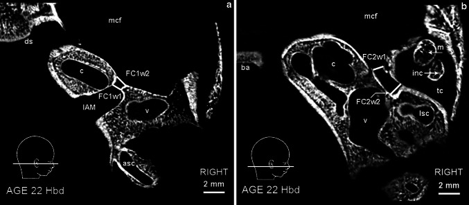
Fig. 3.
Micro-CT scan of the right temporal bone at 22 Hbd (horizontal section). a asc anterior semicircular canal, c cochlea, ds dorsum sellae, mcf middle cranial fossa, v vestibule. FC1w1, FC1w2, IAM—parameters described in the text and Table 1. b ba basilar portion of the occipital bone, c cochlea, inc incus, m malleus, mcf middle cranial fossa, lsc lateral semicircular canal, tc tympanic cavity, v vestibule. FC2w1, FC2w2—parameters described in the text and Table 1

